-Our new joint application note with Proteintech demonstrates how combining FlexAble 2.0 with Magnify Expansion Microscopy accelerates multiplexed super-resolution imaging.
At Magnify Biosciences, we believe that high-resolution imaging should be accessible and efficient. A major part of that vision involves collaborating with other innovators in the field to solve common workflow bottlenecks. We are pleased to announce a new joint application note with our partners at Proteintech, showcasing a workflow that significantly speeds up the antibody staining process for expansion microscopy.
The Challenge: Complex Staining in Thick Tissue
While our universal anchoring system allows for 11x expansion of thick tissue samples, traditional immunofluorescence workflows can be time-consuming. Researchers often face “host species limits” (e.g., requiring different primary antibodies, such as Mouse vs. Rabbit) and long incubation times for secondary antibodies to penetrate expanded gels.
A Collaborative Solution: FlexAble 2.0 + Magnify
By working together, our teams validated a workflow that combines Proteintech’s FlexAble 2.0 Antibody Labeling Kits with our Magnify Expansion Kits. This combination offers a streamlined approach for researchers looking to multiplex without the usual constraints.
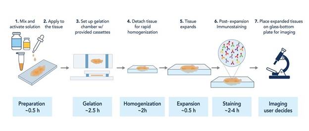
Figure 1. Overview of the Magnify™ Workflow (from the application note).
Key findings from our collaboration include:
In our joint study, we successfully labeled and expanded Tom20 (Mitochondria) and GOLGA2 (Golgi) using the same sample with Rabbit Polyclonal antibodies. The resulting images show distinct ultrastructures resolved at ~25nm with no crosstalk.
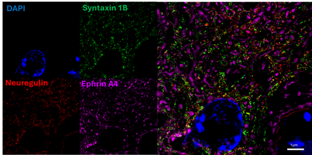
Figure 2. Immunofluorescent images of the mouse brain specimens expanded using a Magnify™ Kit and stained with FlexAble 2.0 Antibody Labeling Kit. Expanded mouse brain tissue was stained with Rabbit anti-Syntaxin 1B (Proteintech, 83298-1-RR), Rabbit anti-Neuregulin (Proteintech, 83323-6-RR), and Rabbit anti-Ephrin A4 (Proteintech, 82951-1-RR). The antibodies were conjugated using the FlexAble 2.0 Antibody Labeling Kit (Syntaxin 1B conjugated with KFA501 CoraLite® Plus 488; Neuregulin conjugated with KFA502 CoraLite® Plus 555; and Ephrin A4 conjugated with KFA503 CoraLite® Plus 647). The sample was imaged using a Nikon Eclipse Ti2 epifluorescence microscope equipped with a CSU-W1 spinning disk confocal module using a CFI Plan Apochromat VC 60×C WI (1.2 NA) objective (from the application note).
We invite you to read the full details of this collaboration and view the imaging results on Proteintech’s website.