We’re thrilled to share that Magnify™ Biosciences’ expansion microscopy technology has been featured in FluoroFinder’s latest article, “Steric Effects in Multiplexed Immunofluorescence.“ This article highlights how expansion microscopy (ExM) provides a powerful solution for overcoming steric hindrance and diffusion challenges in highly multiplexed imaging.
As researchers continue to push the limits of multiplexed immunofluorescence, they often encounter two major obstacles:
1️⃣ Steric hindrance – The physical crowding of antibodies and biomolecules, which can block access to target epitopes.
2️⃣ Limited reagent diffusion – Poor penetration of antibodies in thick tissue samples, reducing signal intensity and imaging depth.
The FluoroFinder article outlines three main approaches to addressing these issues:
✔ Using smaller antibodies (e.g., VHH antibodies)
✔ Expanding the sample itself (Expansion Microscopy)
✔ Employing sequential staining methods
Magnify™ Biosciences’ technology takes center stage in the second category, providing an innovative approach to breaking through steric barriers and improving antibody penetration in thick samples.
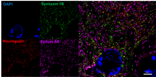
Magnify™ Expansion Microscopy physically separates biomolecules within a hydrogel matrix, allowing antibodies to access previously hidden epitopes. As our CSO Aleks Klimas explains in the article:
“Magnify™ can effectively relieve steric hindrance in antibody binding to different epitopes within protein complexes. The molecular decrowding achieved through expansion physically separates biomolecules within the gel, improving antibody accessibility to previously masked epitopes.”
This innovation unlocks new possibilities in multiplexed imaging, enabling researchers to visualize intricate protein interactions at an unprecedented level.
Antibody penetration in whole mounts and thick tissue sections is another common challenge in structural biology and immunofluorescence imaging. The FluoroFinder article emphasizes how Magnify’s expansion technology dramatically improves diffusion efficiency:
“Another advantage of tissue expansion is improved diffusion of labels into thick samples. Especially for large molecules like antibodies, as the matrix swells, physical obstructions are reduced, increasing the diffusion rate.”
For researchers working with formalin-fixed paraffin-embedded (FFPE) tissues, organoid models, or large biological specimens, Magnify™ Expansion Microscopy enables deep tissue labeling and visualization with greater clarity.
A standout feature of the Magnify™ Expansion Kit is its adjustable expansion factor, which allows researchers to fine-tune sample expansion based on their imaging needs.
🔹 3× to 6× expansion – Achieved by adjusting the salt concentration in the imaging buffer.
🔹 Full 11× expansion – Simply place the gel in pure water for maximum expansion.
This flexibility ensures that Magnify™ Biosciences’ technology seamlessly integrates into diverse imaging workflows, providing customized solutions for every experiment.

📸 Mouse colon FFPE slide before (left) and after Magnify™ expansion, demonstrating adjustable expansion factors.
One of the most exciting aspects of expansion microscopy is its ability to achieve super-resolution-like imaging without requiring expensive new equipment. The article highlights this advantage:
“Optically, ExM enables super-resolution-like image acquisition with traditional, diffraction-limited fluorescent microscopes present in nearly every structural biology lab.”
With Magnify™ Expansion Microscopy, researchers can achieve nanoscale imaging without needing STED, SIM, or PALM/STORM microscopy systems—making super-resolution more accessible and affordable than ever.

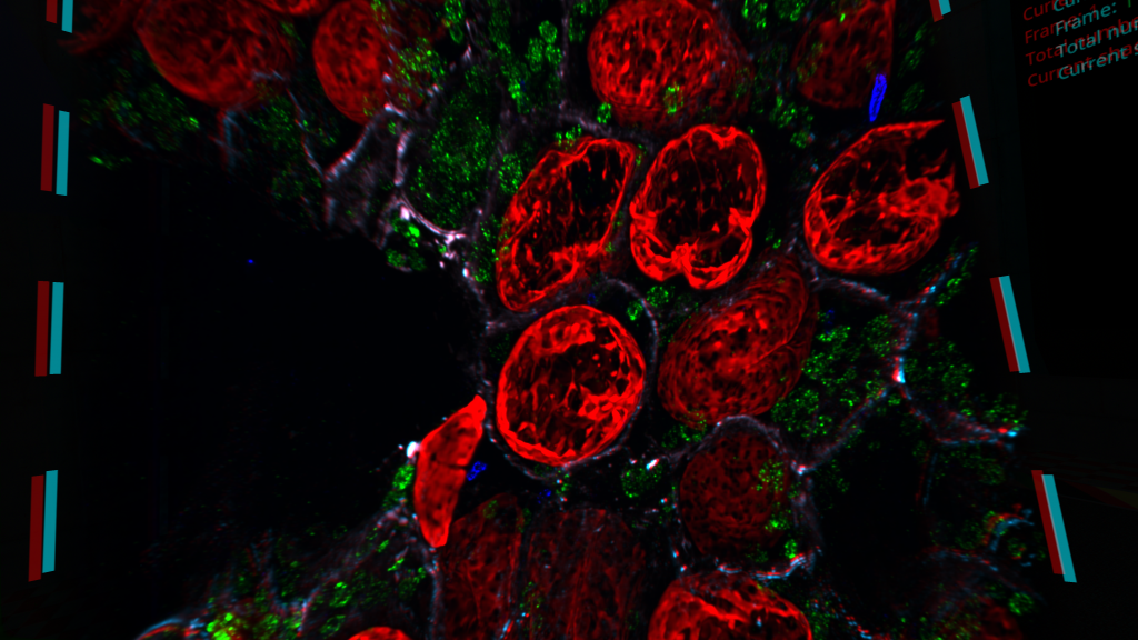
📸 Human platelet cell before (left) and after (right) expansion using the Magnify™ Expansion Kit, revealing detailed subcellular structures.
In the article, Aleks Klimas further explains what makes our expansion microscopy technology unique:
“Magnify™ stands out as a universal, high-expansion, and robust expansion microscopy method that works across diverse sample types and fixation methods while preserving biomolecules and enabling post-expansion staining. This makes it a powerful alternative to existing protocols, especially for applications requiring high-plex imaging in challenging samples like FFPE tissues and infected tissues. Our kits provide validated and tested reagents, ensuring reliable sample expansion without the need for users to source materials independently.”
Unlike other expansion methods, Magnify’s standardized kits provide validated reagents and standardized protocols, ensuring consistent, reproducible expansion microscopy results for researchers.
If you’re encountering steric hindrance or diffusion limitations in your multiplexed immunofluorescence experiments, the Magnify™ Expansion Kit offers a powerful, easy-to-use solution.
🔬 Learn more about our products and applications: Visit our website
📞 Talk to our experts: Contact us to discuss how Magnify™ Biosciences can enhance your imaging workflow.
📖 Read the full article on FluoroFinder’s website: FluoroFinder Blog
Magnify™ Biosciences is pioneering next-generation expansion microscopy solutions, enabling researchers to achieve super-resolution imaging with standard microscopes. Our technology is designed for high-plex imaging across diverse sample types, accelerating discoveries in structural biology, neuroscience, immunology, and infectious disease research.
Follow us for the latest innovations in expansion microscopy!